NEET-XI-Biology
20: Locomotion and Movement
Note: Please signup/signin free to get personalized experience.
Note: Please signup/signin free to get personalized experience.
10 minutes can boost your percentage by 10%
No item to list.
Note: Please signup/signin free to get personalized experience.
- #20 - Locomotion and Movement
- #Section : IPage No 313:
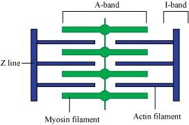
- Qstn #1Draw the diagram of a sarcomere of skeletal muscle showing different regions.
Ans : The diagrammatic representation of a sarcomere is as follows:
- Qstn #2Define sliding filament theory of muscle contraction.
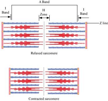
Ans : The sliding filament theory explains the process of muscle contraction during which the thin filaments slide over the thick filaments, which shortens the myofibril.
Each muscle fibre has an alternate light and dark band, which contains a special contractile protein, called actin and myosin respectively. Actin is a thin contractile protein present in the light band and is known as the I-band, whereas myosin is a thick contractile protein present in the dark band and is known as the A-band. There is an elastic fibre called z line that bisects each I-band. The thin filament is firmly anchored to the z line. The central part of the thick filament that is not overlapped by the thin filament is known as the H-zone.
During muscle contraction, the myosin heads or cross bridges come in close contact with the thin filaments. As a result, the thin filaments are pulled towards the middle of the sarcomere. The Z line attached to the actin filaments is also pulled leading to the shortening of the sarcomere. Hence, the length of the band remains constant as its original length and the I-band shortens and the H-zone disappears.
- Qstn #3Describe the important steps in muscle contraction.
Ans : During skeletal muscle contraction, the thick filament slides over the thin filament by a repeated binding and releases myosin along the filament. This whole process occurs in a sequential manner.
Step 1: Muscle contraction is initiated by signals that travel along the axon and reach the neuromuscular junction or motor end plate. Neuromuscular junction is a junction between a neuron and the sarcolemma of the muscle fibre. As a result, Acetylcholine (a neurotransmitter) is released into the synaptic cleft by generating an action potential in sarcolemma.
Step 2: The generation of this action potential releases calcium ions from the sarcoplasmic reticulum in the sarcoplasm.
Step 3: The increased calcium ions in the sarcoplasm leads to the activation of actin sites. Calcium ions bind to the troponin on actin filaments and remove the tropomyosin, wrapped around actin filaments. Hence, active actin sites are exposed and this allows myosin heads to attach to this site.
Step 4: In this stage, the myosin head attaches to the exposed site of actin and forms cross bridges by utilizing energy from ATP hydrolysis. The actin filaments are pulled. As a result, the H-zone reduces. It is at this stage that the contraction of the muscle occurs.
Step 5: After muscle contraction, the myosin head pulls the actin filament and releases ADP along with inorganic phosphate. ATP molecules bind and detach myosin and the cross bridges are broken.
Stage 6: This process of formation and breaking down of cross bridges continues until there is a drop in the stimulus, which causes an increase in calcium. As a result, the concentration of calcium ions decreases, thereby masking the actin filaments and leading to muscle relaxation.
- #4-aActin is present in thin filamentAns : Answer: True
- #4-bH-zone of striated muscle fibre represents both thick and thin filaments.Ans : Answer: False
H -zone of striated muscle fibre is the central part of the thick filament that is not overlapped by the thin filament.
- #4-cHuman skeleton has 206 bones.Ans : Answer: True
- #4-dThere are 11 pairs of ribs in man.Ans : Answer: False
There are 12 pairs of ribs in a man.
- #4-eSternum is present on the ventral side of the body.Ans : Answer: True
- #5-aActin and MyosinAns : Actin and Myosin
Actin
Myosin
1 Actin is a thin contractile protein. 1 Myosin is a thick contractile protein. 2. It is present in light bands and is called an isotropic band. 2 It is present in dark bands and is called an anisotropic band.
- #5-bRed and White musclesAns : Red and White muscles
Red muscle fibre
White muscle fibre
1 Red muscle fibres are thin and smaller in size. 1 White muscle fibres are thick and larger in size. 2 They are red in colour as they contain large amounts of myoglobin. 2 They are white in colour as they contain small amounts of myoglobin 3 They contain numerous mitochondria. 3 They contain less number of mitochondria. 4 They carry out slow and sustained contractions for a long period. 4 They carry out fast work for short duration. 5 They provide energy by aerobic respiration. 5 They provide energy by anaerobic respiration.
- #5-cPectoral and Pelvic girdleAns : Pectoral and Pelvic girdle
Pectoral girdle
Pelvic girdle
1 It is a skeletal support from where the forelimbs of vertebrates are attached. 1 It is a skeletal support form where the hind limbs of vertebrates are attached. 2 It is composed of two
Bones namely, clavicle or collar bones and scapula or shoulder bone.
2 It is composed of three bones, upper ileum, inner pubic, and ischium.