NEET-XI-Biology
18: Body Fluids and Circulation
Note: Please signup/signin free to get personalized experience.
Note: Please signup/signin free to get personalized experience.
10 minutes can boost your percentage by 10%
No item to list.
Note: Please signup/signin free to get personalized experience.
- Qstn #10Sino-atrial node is called the pacemaker of our heart. Why?
Ans : The sino-atrial (SA) node is a specialised bundle of neurons located in the upper part of the right atrium of the heart. The cardiac impulse originating from the SA node triggers a sequence of electrical events in the heart, thereby controlling the sequence of muscle contraction that pumps blood out of the heart. Since the SA node initiates and maintains the rhythmicity of the heart, it is known as the natural pacemaker of the human body.
- Qstn #11What is the significance of atrio-ventricular node and atrio-ventricular bundle in the functioning of heart?
Ans : The atrioventricular (AV) node is present in the right atrium, near the base of the inter-auricular septum that separates the right auricle from the ventricle. It gives rise to the bundle of His that conducts the cardiac impulses from the auricles to the ventricles. As the bundle of His passes the ventricle along the inter-ventricular septum, it divides into two branches - the right ventricle and the left ventricle.
The end branches of this conducting system then forms a network of Purkinje fibres that penetrate into the myocardium. The auricular contraction initiated by the wave of excitation from the sino-atrial node (SA node) stimulates the atrio-ventricular node, thereby leading to the contraction of ventricles through the bundle of His and Purkinje fibres. Hence, the atrio-ventricular node and the atrioventricular bundle play a role in the contraction of ventricles.
- Qstn #12Define a cardiac cycle and the cardiac output.
Ans : Cardiac cycle is defined as the complete cycle of events in the heart from the beginning of one heart beat to the beginning of the next. It comprises three stages - atrial systole, ventricular systole, and complete cardiac diastole.
Cardiac output is defined as the amount of blood pumped out by the ventricles in a minute.
- Qstn #13Explain heart sounds.
Ans : Heart sounds are noises generated by the closing and opening of the heart valves. In a healthy individual, there are two normal heart sounds called lub and dub. Lub is the first heart sound. It is associated with the closure of the tricuspid and bicuspid valves at the beginning of systole. The second heart sound dub is associated with the closure of the semilunar valves at the beginning of diastole.
These sounds provide important information about the condition and working of the heart.
- Qstn #14Draw a standard ECG and explain the different segments in it.
Ans : Electrocardiogram is a graphical representation of the cardiac cycle produced by an electrograph.
The diagrammatic representation of a standard ECG is shown below.
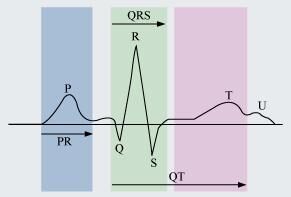
A typical human electrocardiogram has five waves - P, Q, R, S, and T. The P, R, and T-waves are above the base line and are known as positive waves. The Q and S-waves are below the base line and are known as negative waves. The P-wave is of atrial origin, while the Q, R, S, and T-waves are of ventricular origin.
(a) The P-wave indicates atrial depolarisation. During this wave, the impulse of contraction is generated by the SA node. The PQ-wave represents atrial contraction.
(b) The QR-wave is preceded by ventricular contraction. It represents the spread of the impulse of contraction from the AV node to the wall of the ventricle. It leads to ventricular depolarisation.
(c) The RS-wave represents ventricular contraction of about 0.3 sec.
(d) The ST-wave represents ventricular relaxation of about 0.4 sec. During this phase, the ventricles relax and return to their normal state.
(e) The T-wave represents ventricular relaxation.