ICSE-X-Biology
08: The Nervous System
Note: Please signup/signin free to get personalized experience.
Note: Please signup/signin free to get personalized experience.
10 minutes can boost your percentage by 10%
Note: Please signup/signin free to get personalized experience.
- #1-dName the cells of the retina and their respective pigments which get activated (1) in the dark and (2) in the light.digAnsr: IAns : In the dark: Cells - rod cells, Pigment - rhodopsin
In the light: Cells - cone cells, Pigment - iodopsin
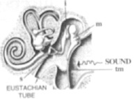
- #2-aGive the technical term for the structure found in the inner ear.Ans : The middle ear or membranous labyrinth has two structures inside it, the cochlea and the semi-circular canals.
- #2-bName the three small bones present in the middle ear. What is the biological term for them collectively?Ans : Malleus, incus and stapes
- #2-cName the part of the ear associated with (1) static balance (2) hearing (3) dynamic balance. (d) Name the nerve, which transmits messages from the ear to the brain.Ans : Static balance - Utriculus and sacculus (inner ear)
Hearing - Internal ear Dynamic balance - Semi-circular canals (inner ear) Collectively they are termed as ossicles.
- #3-aname the corresponding parts of the eye the camera shown here that are comparable in function.Ans : Cornea is comparable to the lens cover of the camera. The iris and pupil act like the aperture of a camera.
- #3-bExplain the mode of working and the functions of the parts of the eye mentioned aboveAns : The cornea is the eye's main focusing element. It takes widely diverging rays of light and bends them through the pupil; the rays are further converged by the lens.
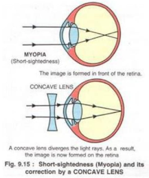
- Qstn #4Given below is a diagram depicting a defect of the human eye. Study the same and answer the questions that follow:
- #4-aName the defect shown in the diagramAns : Myopia
- #4-bGive two possible reasons for this defectAns : The two possible reasons for myopia are either the eye ball is lengthened from front to back or the lens is too curved.
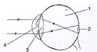
- #4-cName the parts labelled 1 to 4Ans : 1-vitreous humour, 2-blind spot, 3-lens, 4-pupil
- #4-dName the type of lens used to correct this eye defectAns : Concave lens
- #4-eDraw a labelled diagram to show how the above mentioned defect is rectified using the lens named above.Ans :